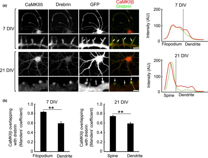
Figure 2.

CaMKIIβ colocalizes with drebrin in cultured hippocampal neurons. (a) CaMKIIβ was localized in dendrites of immature neurons. CaMKIIβ colocalized with drebrin in dendritic filopodia [arrows, 7 days in vitro (DIV)] and dendritic spines of a mature neuron (arrows, 21 DIV). Dashed yellow lines in the merged images of 7 DIV and 21 DIV indicate the positions of the line scan graphs shown right. The line scans show CaMKIIβ (red) and drebrin (green) pixel intensities across the region of interest. AU, Arbitrary units. Scale bars, 30, 2 μm (dendrite images). (b) Quantification of Mander's coefficients for CaMKIIβ overlap with drebrin. Data are presented as means ± SEM (n = 86 filopodia for 7 DIV neuron, n = 29 dendrite regions for 7 DIV neuron, n = 74 spines for 21 DIV neuron, n = 35 dendrite regions for 21 DIV neuron, **p < 0.01, Welch's t‐test).
