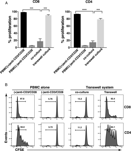
FIGURE 3.
PMN-mediated inhibition of T cell proliferation requires cell-cell contact. Flow cytometric analysis of T cell proliferation in the presence of PMN using a Transwell system. (A) PMN were mixed with CFSE-labeled PBMC at a 4:1 ratio in coculture with anti-CD3/CD28 (coculture). To separate PBMC and PMN, PBMC were cultured in the bottom chamber and PMN were placed in the top chamber of a 96-well Transwell culture plate. Representative results from one of three different donors performed in triplicate are expressed as mean ± SEM. ****p < 0.0001, ***p < 0.01. (B) Representative results of three different donors are shown. Numbers on histograms represent the percentage of proliferating T cells. Fixed PMN were used.

