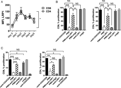
FIGURE 6.
LRP1 blockade prevents PMN-mediated inhibition of T cell proliferation. (A) PBMC from healthy donors were stimulated with anti-CD3/CD28 mAbs for up to 5 d, and LRP1 surface expression was determined by flow cytometry. Graph shows the median fluorescence intensity (MFI) of LRP1 on CD8 and CD4 T cells, respectively. Representative results from one of two different donors performed in duplicates are shown. Error bars represent mean ± SEM. *p < 0.05. (B) Fixed PMN were cocultured with activated PBMC at a ratio of 3:1 for 5 d in the presence or absence of anti-LRP1 mAb (clone 8G1) or mIgG isotype control. Data from three different donors (n = 3) are expressed as mean ± SEM. (C) Fixed PMN were cocultured with sort-purified CD8 or CD4 T cells activated with anti-CD3/CD28 mAbs at 3:1 ratio for 5 d in the presence or absence of anti-LRP1 mAb (clone 8G1) or mIgG isotype control. Results shown as mean ± SEM are representative of three different donors performed in duplicates. ***p < 0.001, **p < 0.01, *p < 0.05, p = NS.

