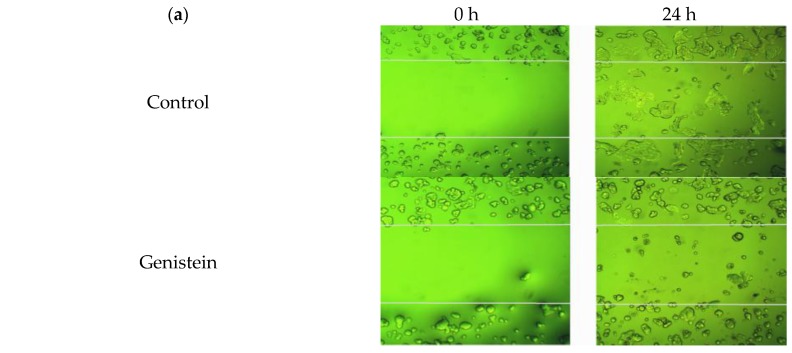
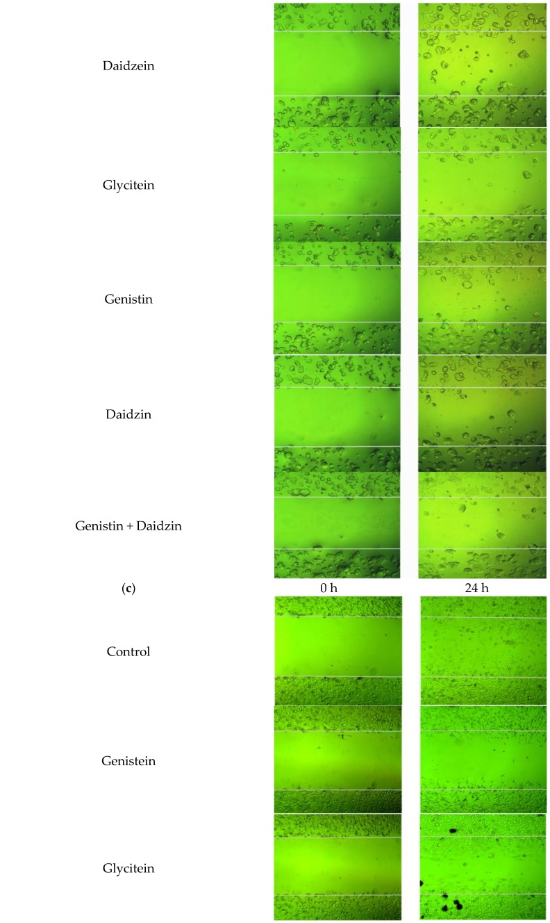
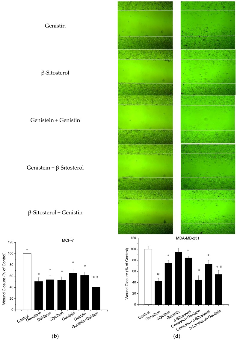
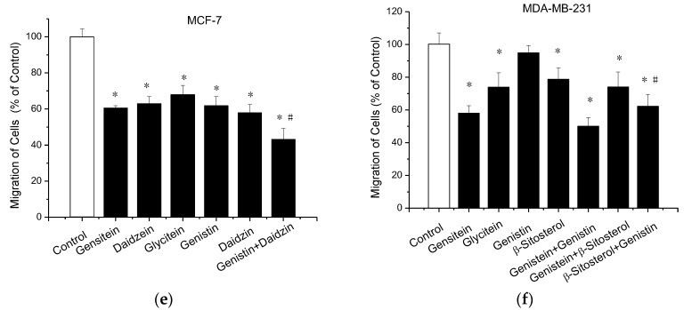
Figure 4.
Inhibition of cell invasion and migration measured by wound healing assay and transwell chamber assay. For wound-healing assay, wounds were made when cells were 90–100% confluent. Overnight, cells were treated with samples, or control. The closure of wounds in MCF-7 cells and MDA-MB-231 cells were imaged (a,c) and quantitatively measured (b,d) at 0 h and 24 h. For transwell chamber assay, MCF-7 (e) and MDA-MB-231 (f) Cells were treated with samples, or control for 48 h. Cells suspended in serum-free medium were seeded on the upper membrane of transwell chamber and incubated for 48 h. Complete growth medium was added on the bottom. Cells on the lower membrane of chambers were counted. Data are presented as mean ± SD. * indicates a significant difference compared to the control (p < 0.05). # indicates a significant difference compared to treatment with samples by single (p < 0.05).


