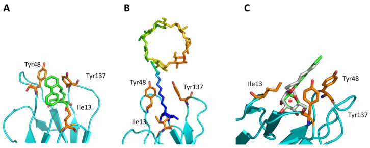
Figure 4.
Recent results including the dynamics aspect of the FimH-ligand complex (formation). (A) A minor MD conformation (populated with 11%) shows the C117 second phenyl ring orientated towards C117 [76]. (B) The position of bCD in the binding pocket (PDB code: 5AB1). The ligand is colored according to the structure factor (from blue: rigid to red: highly flexible). (C) Comparison of the septanoside position (green; HS; PDB code: 5CGB) compared to a mannoside compound (grey; HM; PDB code: 4BUQ). The difference in the sugar ring is highlighted by a red asterisk.

