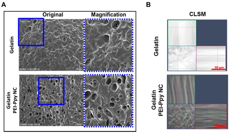
Figure 3.
(A) Morphology and porous structure of the test sample detected by SEM: PEI–Ppy‒NC-loaded gelatin hydrogel (Gelatin, scale bar = 500 μm; PEI–Ppy‒NC-loaded gelatin, scale bar = 1 mm); (B) representative X–Z plane confocal laser scanning microscopy (CLSM) image of gelatin and gelatin containing PEI‒Ppy‒NC, showing the distribution of dark nanoparticles inside the gelatin hydrogel matrix.

