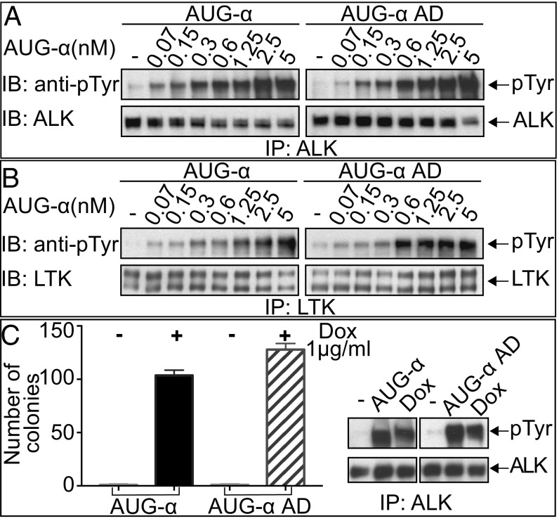
Fig. 2.
Deletion of the N-terminal variable region of AUG-α does not affect kinase activity of ALK or LTK. (A and B) Immunoblot analysis of ALK (A) or LTK (B) autophosphorylation stimulated by different concentrations (as indicated) of purified AUG-α or AUG-α AD. NIH 3T3 cells stably expressing ALK or LTK were stimulated with increasing concentration of AUG-α or AUG-α AD for 10 min at 37 °C. Lysates of unstimulated or AUG-α–stimulated cells were subjected to immunoprecipitation (IP) using anti-ALK or anti-LTK antibodies followed by SDS/PAGE and immunoblotting (IB) with anti-pTyr (IB: pTyr) or anti-ALK or anti-LTK antibodies (as indicated). (C) NIH 3T3 cells were stably transfected with ALK and a Dox-inducible construct of AUG-α or AUG-α AD. (Right) Immunoblot analysis of ALK autophosphorylation stimulated by DMSO (indicated as by “−”) and 50 μg of purified AUG-α or AUG-α AD, or Dox. (Left) Soft agar colony formation assay. Double-stable NIH 3T3 cells were treated with either DMSO or 1 µg/mL of Dox and grown in soft agar for 2 wk. Colonies were stained with crystal violet, counted, and plotted. Each experiment was performed in triplicate, and SD was calculated and plotted for each experiment.

