Fig. 3.
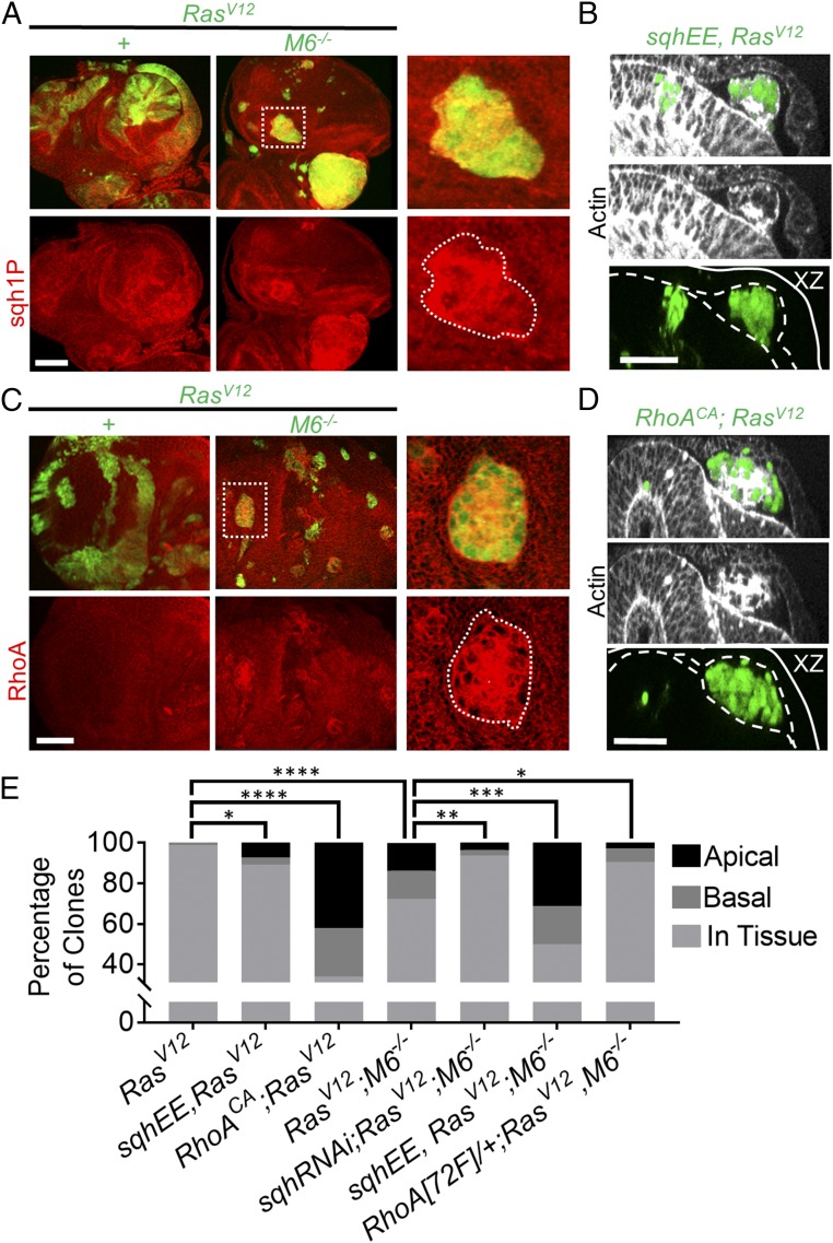
RasV12; M6−/− clones apically delaminate in a RhoA-activated myosin-dependent manner. (A) RasV12 and RasV12; M6−/− eye-disk clones were stained for sqh1P to indicate active myosin. (Scale bar: 50 um.) n = 5. (Right) Boxed area enlarged. (B) sqhEE; RasV12 eye-disk clones. Dashed lines represent the apical domains of the peripodial membrane (top dashed line) and the disk proper (bottom dashed line), and the solid line represents the basal surfaces. (Scale bar: 20 um.) (C) RasV12 and RasV12; M6−/− eye-disk clones were stained for RhoA (n = 5). (Scale bar: 50 um.) (Right) Boxed area enlarged. (D) RhoACA was expressed in RasV12 eye-disk clones and eye discs examined for apical delamination. Dashed lines represent the apical surfaces of the periopodial membrane and disk proper, and the solid line represents the basal surfaces. (Scale bar: 20 um.) (E) Quantification of the localization of clones of indicated genotypes. (n = 84 clones for RasV12; 87 clones for RasV12; M6−/−; 83 clones for sqhEE, RasV12; 112 clones for sqhRNAi; RasV12; M6−/−; 122 clones for sqhEE, RasV12; M6−/−; 50 clones for RhoACA, RasV12; and 106 clones for Rho[72F]/+; RasV12, M6−/−) Statistical significance was analyzed using χ2 analysis with each genotype being compared with the relevant control. *P < 0.05, **P < 0.01, ***P < 0.001, and ****P < 0.0001.

