Fig. 1.
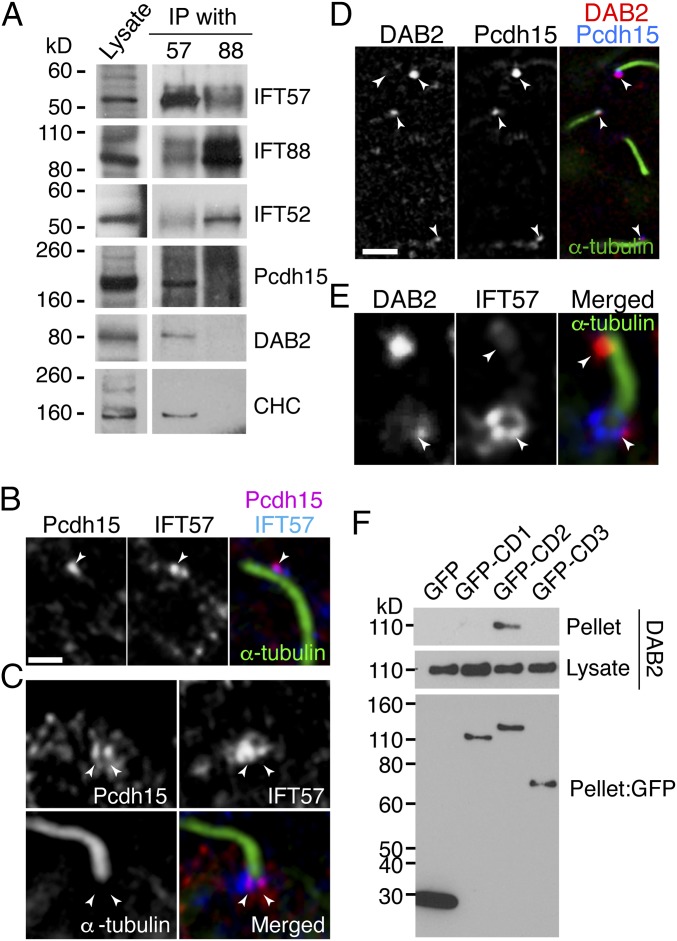
IFT components bind Pcdh15 in kinocilia. (A) IP assay from E10 chick cochlear lysate with anti-IFT57 or anti-IFT88 antibodies. Pcdh15 is cosedimented with IFT57 together with CHC and DAB2. (B) Pcdh15 (magenta) and IFT57 (blue) are colocalized in kinocilia (green, stained with anti–α-tubulin antibody) of chick cochlea (BP) at E10 (arrowhead). (C) Pcdh15 (red) was colocalized with IFT57 (blue) in the base of kinocilia stained with α-tubulin (green). (D) DAB2 (red) and IFT57 (blue) are also colocalized in kinocilia (green) and at the base of the kinocilia. (E) DAB2 (magenta) and Pcdh15 (blue) are colocalized in kinocilia (arrowheads) of E10 chick basilar papilla. (Scale bars: 5 µm in A, C, and D; 10 µm in E.) (F) The GFP-tagged Pcdh15 isoform constructs depicted were coexpressed with DAB2 in BMT10 cells. IP with anti-GFP antibody followed by a DAB2 Western blot shows DAB2 specifically cosediments with the GFP-CD2 domain, but not CD1 or CD3. A GFP Western blot shows similar expression for all tagged isoforms in cells.

