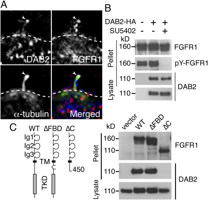
Fig. 6.
DAB2 binds FGFR1 independently of FGFR1 activity. (A) DAB2 (red) and FGFR1 (blue) were colocalized in the tip of kinocilia (arrowheads). (B) IP of DAB2-HA pulled down FGFR1, independently of FGFR1 activity, in overexpressing BMT-10 cells. (C) FGFR1 mutant construct, lacking the FRS-binding motif (∆FBD construct), was still able to bind DAB2 at the same levels as in WT FGFR1 in an in vitro binding assay. An FGFR1 construct lacking the intracellular domain (∆C construct) was unable to bind DAB2, however.

