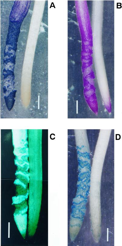
Figure 1.
Histochemical detection of lipid peroxidation and other events caused by aluminum in pea roots. Pea seedlings were treated with (left) or without (right) 10 μm aluminum in 100 μm CaCl2 (pH 4.75) for 24 h. The roots were stained with either hematoxylin (A, aluminum accumulation), Schiff's reagent (B, lipid peroxidation), aniline blue (C, callose production), or Evans blue (D, the loss of plasma membrane integrity; see “Materials and Methods”). The positive staining of each technique in the photomicrographs shows as bright images in panels (A, B, and D) and as a fluorescent image in panel (C). Bar in each graph indicates 1 mm.

