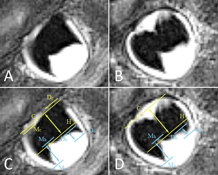
Figure 3.

(A, B) Lower right mandibular third molar depicted on two consecutive MRI slices. Slice (A) is situated more buccally than slice (B). The pulp chamber has a trapezoidal shape, corresponding to stage 3. To exclude stage 4, MR crown height and MR root length have to be evaluated as illustrated in images (C, D). (C) Copy of image (A), with marked landmarks and distances to consider in order to allocate a stage. Lines are perpendicular to the tooth axis. Distances (arrows) are evaluated along the tooth axis. Line Dc is at the distal cusp tip, while Mc is at the mesial cusp tip. Line C represents the midpoint between the distal and mesial cusp tips. Line Dh is at the distal pulp horn while line Mh is at the mesial pulp horn. Line H represents the midpoint between the distal and mesial pulp horns. Line Da is at the most apical point of the distal root. Line Ma is at the most apical point of the mesial root. The distance between lines C and H is the MR crown height based on this slice. The distance between lines Dh and Da is the distal MR root length, while the distance between lines Mh and Ma is the mesial MR root length. (D) Copy of image (B). Both cusp tips are at the same level on this slice, represented by line C. MR crown height is larger than on the previous image, whereas the distal MR root length is smaller. To allocate a stage, MR crown height on image (D) is the most appropriate, while the distal MR root length on image (C) is the most appropriate. Because the third molar is tilted bucco-lingually, the observer has to scroll through consecutive slices to decide on the most appropriate measures to consider. In slice (C) the crown is transsected more buccally, so part of the crown is not depicted. By contrast, the distal root apex is situated more buccally than the crown, so it is best depicted in slice (D). Because the distal root is shorter than MR crown height, this third molar is in stage 3.
