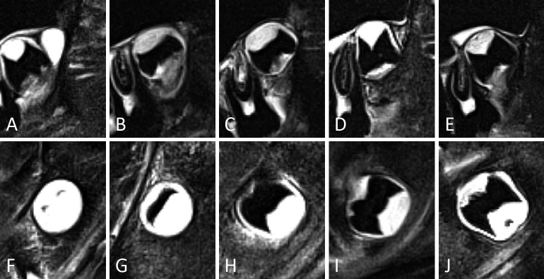
Figure 6.
Representative examples of third molars in developmental stages 0 to 3, in the upper (A-E) and lower jaw (F-J). For some stages different appearances are illustrated. (A) Stage 0. The crypt of the third molar shows no calcification. It is seen as a clearly delineated white area. (F) Stage 1. Cusp tips are seen as separate black areas within the crypt. (B, G) Early stage 2. Cusps are fused. The roof of the pulp chamber is quite flat. (C, H) Late stage 2. The roof of the pulp chamber is more curved than in (B, G). Notice that in (H), the mesial side of the pulp chamber is more mature than the distal side. Thus, for staging the distal side should be considered. The distinction between early and late stage 2 was considered too subjective to consider them as separate stages. (D, I) Stage 3. Notice the pointy appearance of the pulp horns. No furcation was present. (E, J) Stage 3. Notice the furcation. In (J) the distal pulp horn appears curved on this sagittal slice. However, scrolling through the slices and including the coronal slices in the assessment, it was clear that both pulp horns were pointy, like an umbrella top.

