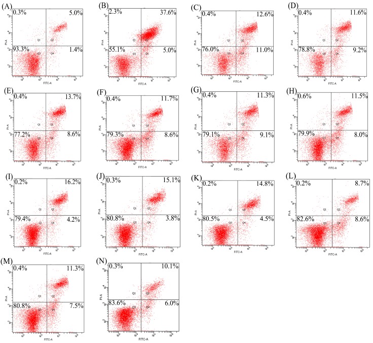
Figure 2.
Detection of apoptotic prevention in the osteoblasts by flow cytomety. (A) control cells; (B) EP-treated cells; (C–H) the cells firstly treated by respective PAH1, PAH3, PNH1, PNH3, PPH1, and PPH3 for 48 h, and then treated by EP for 24 h; (I–N) the cells firstly treated by respective TAH1, TAH3, TNH1, TNH3, TPH1, and TPH3 for 48 h, and then treated by EP for 24 h.

