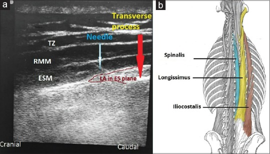
Figure 1.

(a) ESP block given under US guidance at T4 level. Image also shows local anaesthetic in ESP with the muscle getting pushed above (ESM: erector spinae muscle, RMM: rhomboidus major muscle, TZ: trapezius muscle). (b) The three muscles: iliocostalis, longissimus, and spinalis, which forms the ESM along with its attachment to the vertebral column and other bones (Image source: The image has been submitted after permission from Dr. Oliver Jones from the site: http://teachmeanatomy.info/)
