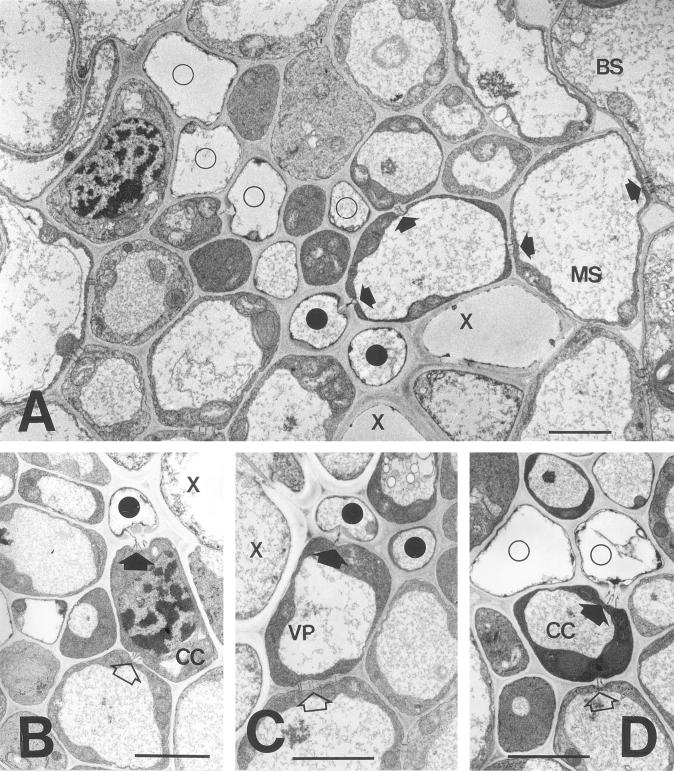
Figure 1.
Electron micrographs of intermediate vascular bundles in a sink barley leaf. A, Overview of vascular bundle. Two thick-walled SEs (black circles) occur adjacent to xylem elements (X). Four thin-walled SEs (white circles) and associated CCs are also shown. A complete symplastic pathway (darts) is evident between the upper thick-walled SE and the bundle sheath (BS). Scale = 10 μm. B, Pore-plasmodesma connection between a thick-walled SE and CC (dart). Note that the CC is also connected to a vascular parenchyma element by branched plasmodesmata (white dart). Scale = 10 μm. C, Plasmodesmatal connections between a thick-walled SE and vascular parenchyma element (dart). The parenchyma element is connected to a neighboring cell by simple plasmodesmata (white darts). Scale = 10 μm. D, Pore-plasmodesma connection between a thin-walled SE and its CC (dart). The CC is in turn connected to a vascular parenchyma element (white dart). Scale = 10 μm.

