Figure 3.
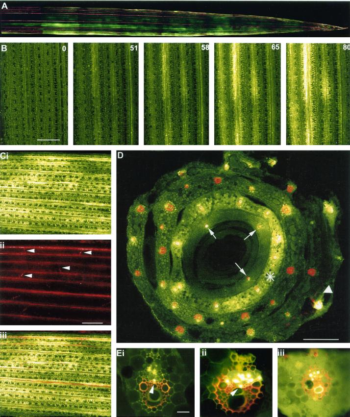
Unloading of CF in sink barley leaves. A, CLSM image of CF unloading in the apical region of a sink leaf of barley. Note the discontinuous exit of dye from the major vascular bundles. The functional xylem of the leaf was labeled with Texas Red dextran. The sink leaf was 5 cm long. B, CLSM imaging of the progression of CF unloading into a sink leaf of barley. The CF was first detected above background at 51 min after labeling of a source leaf. The numbers in the right-hand corner of the remaining images depict the time(s) of subsequent images of the unloading process. Note the rapidity of phloem unloading following the arrival of the dye in the leaf. A discontinuous pattern of dye unloading is also evident. Time in minutes. Scale = 1.0 mm. C, Sink leaf after unloading CF for 100 min. i, Extensive dye spread is evident from the major longitudinal bundles into the mesophyll. ii, Same region of leaf showing functional xylem stained with Texas red dextran. Lateral veins (darts) are evident. iii, Image merge of i and ii. Scale = 1.0 mm. D, CLSM image of the base of a leaf sheath following labeling of a single source leaf (white dart) with CF. Note that in the source leaf, CF is restricted to the vascular bundles. The leaf was also labeled with Texas red to depict functional xylem elements. The bulk of phloem unloading occurs from an inner sink leaf (asterisk) and dye has clearly entered the leaf mesophyll. In the innermost (immature) sink leaf dye is restricted to only three translocating vascular bundles (arrows) due to immaturity of the phloem in this leaf. Scale = 0.5 mm. E, CLSM images of vascular bundles translocating CF. In some of the vascular bundles (i and ii) labeled phloem elements occupy the position expected for thick-walled SEs (darts). iii, Vascular bundle from an unloading sink leaf. Note the presence of dye in the mesophyll. Some CF also remains present within SEs of the phloem. The functional xylem was labeled with Texas red dextran. Scale = 150 μm.

