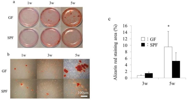
Figure 4.
Calcification in cultured primary osteoblasts from germ-free (GF) and specific pathogen-free (SPF) mice. Calvariae from 8-week-old GF and SPF male mice were harvested, and primary osteoblasts were isolated and cultured in osteoblastic differentiation media. (a) Alizarin red staining of osteoblasts from GF and SPF calvariae cultured for 1, 3, and 5 weeks. Panel (b) shows high-magnification fields of panel (a). (c) The alizarin red–positive area was calculated using a computerized imaging system (ZEN, ZEISS, Oberkochen, Germany). The alizarin red–positive area at 5 weeks was significantly greater in SPF mice than GF mice. Mean values (±SD) are shown * p < 0.05, t-test (n = 4/group). Scale bar = 100 μm (b).

