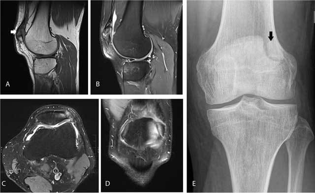
Figure 1.

Symptomatic bipartite patella.
Sagittal T1-WI (A) shows a separate bone fragment at the superolateral aspect of the patella (arrow). Sagittal (B), axial (C) and coronal (D) FS T2-WI reveal bone marrow edema within the accessory fragment and the adjacent superolateral aspect of the patella. Plain radiography (E) confirms a sclerotic delineated accessory fragment at the superolateral aspect of the patella (arrow).
