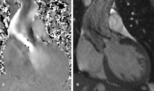
Figure 3A and B.

(A) Coronal flow velocity encoded images show high velocity blood flow across the aortic valve (arrowheads) compatible with a moderate aortic valve stenosis; a peak systolic velocity of 328 cm/s was measured. (B) Coronal cine MRI images of the left ventricular outlet during systole demonstrate a narrowed aortic valve orifice (arrowheads) during systole because of concurrent aortic stenosis.
