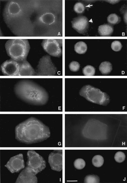
Figure 1.
Localization of Rop GTPase to an endomembrane system in pea tapetal cells. Pea anthers prior to anthesis were collected and squashed as described in text. Squashed anthers were stained with affinity-purified anti-Rop1Ps antibodies (A, C, E, F, and G) or anti-BiP antibodies (I) and an anti-rabbit secondary antibody conjugated with fluorescein. Cells shown in A, C, and I were counterstained with DAPI to reveal the localization of the nucleus (B, D, and J), respectively. Arrow in B indicates the location of the nucleus in tapetal cells, whereas arrowhead indicates the nucleus of microspore mother cells. Negative control is shown in H, in which the primary anti-Rop1Ps antibody was replaced with a pre-immune serum. Bar = 25 μm.

