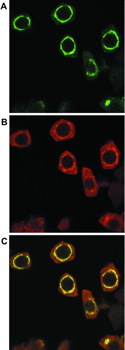
Figure 3.
Colocalization of Rop with the vacuolar annexin VCaB42. Squashed anthers were co-incubated with the rabbit anti-Rop1Ps antibody and mouse anti-VCaB42 antibody. The reaction was first treated with FITC-conjugated anti-rabbit IgG secondary antibodies and then with the Texas Red-conjugated anti-mouse IgG sheep F(ab)2 fragment. After washes, the samples were observed under a laser scanning confocal microscope as described in text. A, Localization of Rop as revealed by FITC fluorescence. B, Localization of VCaB42 as revealed by Texas Red fluorescence. C, Overlay between A and B.

