Figure 5.
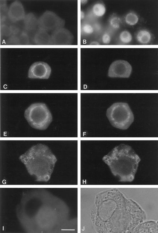
The dynamic localization of Rop to the tonoplast is correlated with vacuole development in pea tapetal cells. Pea anthers from different developmental stages were squashed and co-stained with anti-Rop1Ps (A, C, E, G, and I) and anti-VCaB42 (D, F, and H) antibodies as described in Figure 3. Stained cells were examined under an epifluorescence microscope. At the stage of microsporogenic cells (giving rise to microspore mother cells) and parietal cells (giving rise to the tapetum), only diffuse cytoplasmic staining (A) for Rop is found. At this stage, it is difficult to distinguish the two cell types either by cell shape (A) or nuclear morphology (B). Localization to the tonoplast of tapetal cells (C) appears at the stage of microspore mother cells, whereas little tonoplast localization is observed for VCaB42 (D) at this stage. The tonoplast localization of Rop persists through the early and late meiosis I (E and G, respectively), during which VCaB42 is colocalized with Rop (F and H). By early tetrad stage, Rop staining (I) completely disappears from the tonoplast of the large mature vacuole (see differential interference contrast image in J), whereas VCaB42 staining in the tonoplast remains (data not shown).

