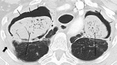
Figure 15.

Aspergilloma. A 58-year-old male sarcoidosis patient also had known long-standing fibrosis. Routine CXR revealed opacities in the apical segments of both lungs. CT showed large content known fibrotic cysts apically with crescent-shaped air (short arrows) anteriorly due to large formed fungus balls (long arrows). There was local pleural thickening (thick arrow) (no symptoms).
