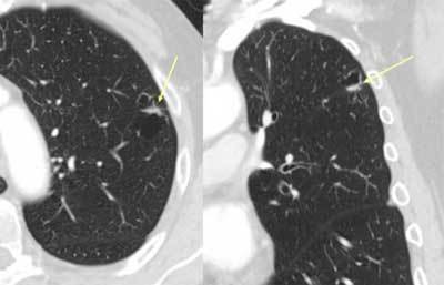
Figure 20.

Lung cancer. A 70-year-old male patient was admitted due to dyspnea when supine, no fever or cough. In the left upper lobe, a mostly thin-walled multicystic lesion is seen, with a short thicker wall in-between (arrows). A biopsy proved adenocarcinoma.
