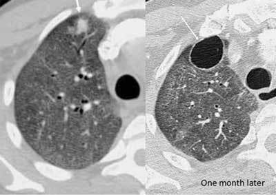
Figure 24.

Pneumocystis jirovecii infection. A 40-year-old male was admitted with acute dyspnea. An initial CT was done for suspected PE. It showed extensive ground glass opacities in both lungs and a small consolidation in the right upper lobe. The patient was subsequently diagnosed with acquired immunodeficiency. One month later (HRCT) the ground glass is less extensive, and the nodule has developed into a thin-walled cyst.
