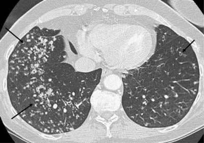
Figure 10.

Miliary tuberculosis. A 65-year-old female with dyspnea was admitted as her pneumonia was not improving despite treatment. The CT was performed with a suspicion of pulmonary embolism. The images show multiple small cavitary nodules in the upper and lower lobes on both sides. Microbiology proved positive for tuberculosis.
