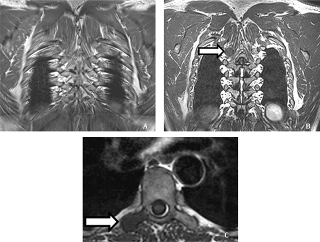
Figure 6.

Detection of a BM in a 63 year old PCa patient. The lesion was missed by both readers on 2DT1 sequence (A) but correctly identified by both on 3D T1 sequence (B). Axial reconstruction obtained with multiplanar reformation at same level confirms the presence of a BM (C). Reprinted from ‘Whole-body 3D T1-weighted MR imaging in patients with prostate cancer: feasibility and evaluation in screening for metastatic disease’ Pasoglou V., Michoux N., Peeters F., Larbi A., Tombal B. Omoumi P., Vande Berg B., Lecouvet F.E, Radiology. 2015 Apr; 275(1): 155–66. [43] With the permission of RSNA.
