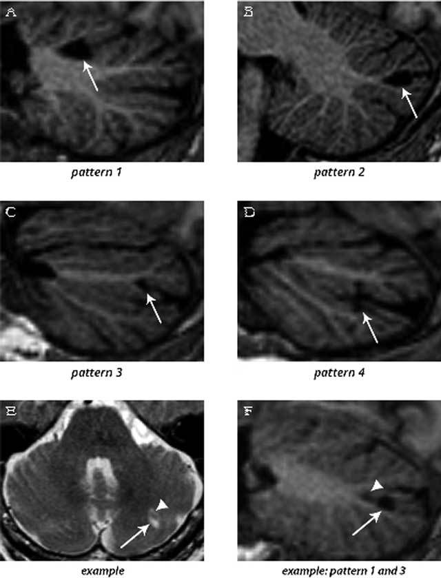
Figure 3.

3D T1-weighted images (sagittal reconstructions) of the brain (A–D and F), providing excellent contrast between gray matter and white matter, show the four patterns by which cerebellar infarcts (arrows) typically affect the cerebellar cortex. An example of two cerebellar infarcts adjacent to each other (E; arrow and arrowhead) are detected on transverse T2WI. Sagittal reconstruction of 3D T1WI of the same two infarcts (F) shows how the topography of one infarct corresponds to pattern 3 (arrow), while the other infarct (arrowhead) corresponds to a small pattern 1 infarct. Notice the sparing of the subcortical and deep white matter in each infarct. The images are cropped to only display the cerebellum. Reproduced from Neuroimage: Clinical [5].
