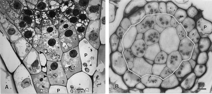
Figure 1.
Longitudinal (A) and cross (B) sections through 5-d-old tobacco root tips grown vertically but placed horizontally for approximately 2 min during the mounting of the samples for high pressure freezing. A, Electron micrographs depicting a meristematic (M) columella cell initial, and three stories of derived columella cells (nos. 1, 2, and 3). Most of the columella cell amyloplasts (arrowheads) appear sedimented toward the lower, distal cell wall. B, Light micrograph of a root tip cross section at the level of the second tier columella cells (cells demarcated by the white line). Due to the offset organization of the columella cells (see asterisks), the section includes the amyloplast-containing layer (arrowheads) of some cells but not others. The columella cells are surrounded by one or two layers of vacuolated peripheral (P) cells. Bar = 5 μm.

