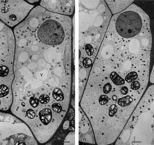
Figure 8.
Effects of drugs that disrupt microfilaments (A) and microtubules (B) on the distribution and organization of nodal ER domains (arrows) in flanking file columella cells. In A, the 1-h treatment with 1 μm latrunculin A is seen to lead to much larger nodal ER domains (see also Figs. 5C and 9B) along the lateral walls, but the number of these domains decreases (data not shown). Note also the proliferation of rough ER membrane sheets (asterisk) between the nucleus and the adjacent upper cell wall. The cell in B was treated with 10 μm propyzamide for 1 h. Only small fragments of what might have been former nodal ER domains (arrows) are seen. N, Nucleus; Am, amyloplast; V, vacuole. Higher magnification views of the nodal ER domains of these samples are shown in Figure 9. Bars = 2 μm.

