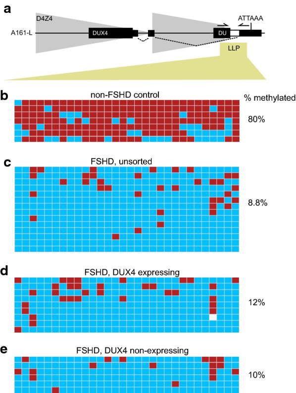
Fig. 2.

CpG methylation density from DUX4 expressing and DUX4 non-expressing arrays in FSHD and non-FSHD control myocytes. CpG methylation events are shown from sorted populations of bisulfite-treated non-FSHD control or FSHD-affected differentiated myocytes. Primers that uniquely amplify the 4qA-161-L haplotype were used so only methylation events from the 4q A161-L haplotype are shown. a Diagram of a full D4Z4 unit and terminal D4Z4 partial unit (LLP) with portions of full length (DUX4) or partial (DU) DUX4 genes shown as black rectangles. The position of LLP PCR primers are shown as converging arrows. b CpG methylation pattern in non-FSHD control myocytes containing a single LLP D4Z4 array (Fig. 1, 2081). c CpG methylation pattern of the LLP region in unsorted FSHD-affected differentiated myocytes (Fig. 1, 2349). d CpG methylation pattern of the LLP region in the DUX4 expressing population of cells from FSHD-affected differentiated myocytes (Fig. 1, 2349). e CpG methylation pattern of the LLP region in the DUX4 non-expressing population of cells from FSHD-affected differentiated myocytes (Fig. 1, 2349). The percentage of CpG methylation are indicated to the right-hand side for each group. The location of methylated cytosines is shown as red squares, and the location of unmethylated cytosines are shown as blue squares. DNA variants which result in a sequence but are no longer a CpG are colored white
