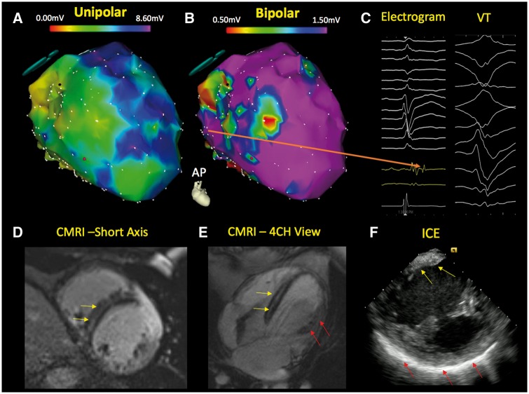
Figure 4.
Electrophysiologic imaging of a non-ischaemic cardiomyopathy patient with ventricular tachycardia. The electroanatomical map of the left ventricle demonstrates a significantly abnormal endocardial unipolar voltage map (A—modified antero-posterior view) and a limited bipolar abnormality (B) in the septum. The unipolar electroanatomic map can provide insight regarding the presence of intramyocardial scar when the bipolar voltage map is relatively normal. Late potentials are also observed in this region during sinus rhythm (C—orange arrow). Cardiac magnetic resonance imaging demonstrates mid myocardial septal scar (yellow arrow) in the short axis (D) and four-chamber view (E). The patient’s intracardiac echocardiography (F) depicts regions of brightness in the septum (yellow arrow) and lateral wall (red arrow). CMRI, cardiac magnetic resonance imaging; ICE, intracardiac echocardiography; VT, ventricular tachycardia.

