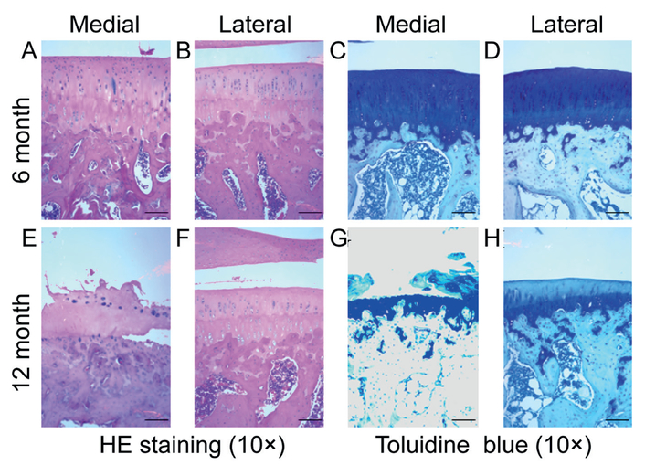
Figure 1.
Histological analysis of 6 and 12 months cartilage. HE and toludine blue staining was used to assess chondrocyte and proteoglycan loss. (A-B): There is minimal cell and matrix disruption in the medial and lateral plateau of the 6 months cartilage. (E-H): In the medial plateau of the 12 months cartilage, there is a cartilage destruction with the crack between calcified and uncalcified cartilage interface, chondrocyte loss, and matrix breakdown with loss of proteoglycans. Even with the intact articular surface, there is a loss of chondrocytes and matrix components in the lateral plateau of 12 month samples. Scale bar = 200 μm.

