Fig. 5.
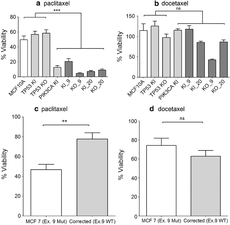
PIK3CA mutations sensitize cells to paclitaxel. Cells were seeded and analyzed as described in Sect. “Materials and methods”. Results shown are representative examples of multiple independent assays. a Cells were maintained in 2.5 nM paclitaxel in assay media for 1 week (***p < 0.001). b Cells were maintained with 60 pM of docetaxel in assay media for 1 week (ns = not significant). MCF10A = parental cells, TP53 KI = MCF10A with TP53 R248W knockin mutation, TP53 KO = MCF10A with TP53 knockout, PIK3CA KI = MCF10A with single PIK3CA E545K knockin, KI_9 = MCF10A with TP53 R248W knockin and PIK3CA E545K knockin, KI_20 = MCF10A with TP53 R248W knockin and PIK3CA H1047R knockin, KO_9 = MCF10A with TP53 knockout and PIK3CA E545K knockin, and KO_20 = MCF10A with TP53 knockout and PIK3CA H1047R knockin. c Parental MCF7 (Ex. 9 MUT) cells with endogenous mutant PIK3CA E545K and Corrected (Ex.9 WT) MCF7 cells engineered with wild-type-only PIK3CA were maintained in 1.2 nM paclitaxel in assay media for 1 week (**p < 0.01). d Parental MCF7 (Ex. 9 MUT) cells with endogenous mutant PIK3CA E545K and Corrected (Ex.9 WT) MCF7 cells engineered with wild-type-only PIK3CA were maintained in 25 pM of docetaxel in assay media for 1 week (ns = not significant)

