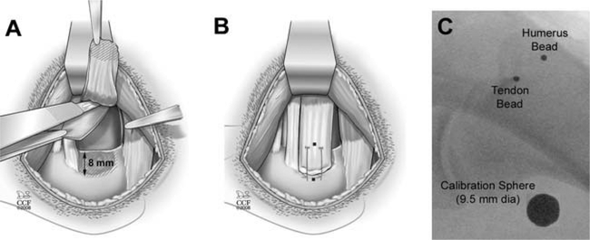
Figure 1.
The canine injury model: (A) A superior two-thirds injury of the infraspinatus tendon was created and a portion of underlying joint capsule was resected. (B) The tendon was repaired to bone using two transosseous sutures. Tantalum beads (shown as black dot ●) were sewn onto the surface of the infraspinatus tendon and implanted into the humerus at the repair site. (C) Intra-operative X-ray shows the tendon bead, bone bead, and calibration sphere (large circle).

