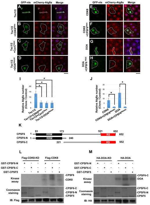
Figure 4. Genetic and biochemical interactions between TOR, CDK8/CycC, DOA, and CPSF6.
(A–J) Genetic interactions between TOR, CDK8/CycC, DOA, and CPSF6. Clonal expression of TSC1 and TSC2 reduced cell size and increased mCherry-ATG8a puncta formation in the larval fat body under fed condition (A), and these effects were suppressed by depletion of CDK8 (B), CycC (C), or DOA (D). Clonal expression of CDK8 (E) or DOA (G) induced mCherry-ATG8a puncta formation under fed conditions, and depletion of CPSF6 inhibits the CDK8 or DOA-induced effects (F and H). Fat body cells were stained with DAPI. Scale bar, 20 μm. (I–J) Quantification of the relative number of mCherry-ATG8a dots per cell. (One-Way ANOVA followed by Bonferroni’s post hoc test; data are represented as mean ± SEM; *P<0.05) (K) Schematic representation of the domain structures of CPSF6 and deletion mutants. (L–M) CDK8 and DOA directly phosphorylate CPSF6 in vitro. Flag-CDK8, CDK8-KD (L), HA-DOA, or HA-DOA-KD (kinase-dead) (M) immunoprecipitated from lysates of transfected cells was used to phosphorylate bacterially expressed recombinant CPSF6-N, CPSF6-C, and CPSF5 in an in vitro kinase assay. Lower panels demonstrate the equal input of GST-fusion proteins and CDK8 or DOA immunoprecipitates.

