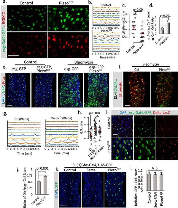
Extended Data Figure 6. Piezo over-expression increases cytosolic Ca2+ level which further triggers proliferation of ISCs but not EBs.

a. Overexpression of PiezoGFP in esg+ cells (esg>Gal4/UAS-PiezoGFP; UAS-RGECO) at 32°C causes an increase in cytosolic Ca2+ (indicated by the calcium reporter RGECO) compared to control (esg>Gal4/UAS-GFP; UAS-RGECO). Representative images from three short time-lapse imaging of cultured fly midguts were shown. Scale bar: 50 μm. b. Typical traces of Ca2+ oscillations in esg+ cells of midgut from either control or PiezoGFP flies from three independent replicates. c. Ca2+ oscillation frequency of esg+ cells from either control or PiezoGFP midguts. Data of 27 cells from three replicates for each condition were shown. d. Statistics for average RGECO signal intensity in all GFP+ cells (blue) and percentage of Ca2+ positive cells (signal higher than 3X s.d. of background) compared to total GFP+ cells (orange). Signal intensities were calculated from 10,000 μm2 regions: n=17 (control), n=22 (PiezoGFP) from three independent experiments. e, Bleomycin (Bleo, 10ug/ml) (5 days treatment) triggers a significant increase of esg+ cell and EE cells in both WT and PiezoKO flies. Represented images from three independent replicates were shown. f, Images of live midguts from WT and PiezoKO flies. Both flies were fed on food containing Bleomycin for 3 days before imaging. g,h. Traces of Ca2+ oscillations in Dl+ stem cells from WT and Piezo mutant flies fed on Bleomycin for 4–5 days. Bleomycin treatment causes some stem cells to maintain constant high Ca2+ levels, while others show reduced oscillation frequency but increased average GCaMP/RFP intensity ratio (G/R ratio). These data show that tissue damage by Bleomycin triggers stem cell proliferation, EE production, and an increase of cytosolic Ca2+, independent of Piezo. 30 cells from n=4 (control), n=4 (Bleo+), and n=5 (PiezoKO + Bleo+) independent guts were plotted. i. Overexpression of PiezoGFP in esg+ cells (32°C) increases the ratio of Dl+ cells (labeled by Dl-lacZ) within the esg+ population. j. Piezo overexpression promotes Dl+ stem cells ratio in esg+ cells. Ratio between Dl+ and esg+ cells within 10,000 μm2 regions: n=21 (control) and n=22 (PiezoGFP) from two independent replicates, are analyzed. k,l. Overexpressing Piezo or knocking down Serca in Su(H)Gbe+ EB cells showed no significant phenotype, suggesting that their effect may be blocked by high Notch activity. Number of midgut areas quantified: n=18 (control), n=20 (Serca-i), n=16 (PiezoGFP). Data are expressed as mean + s.e.m. values. P-values are calculated from two-tailed Student t-test with unequal variance. Scale bar: a,e,f, 50 μm; i, 20 μm; k, 50 μm.
