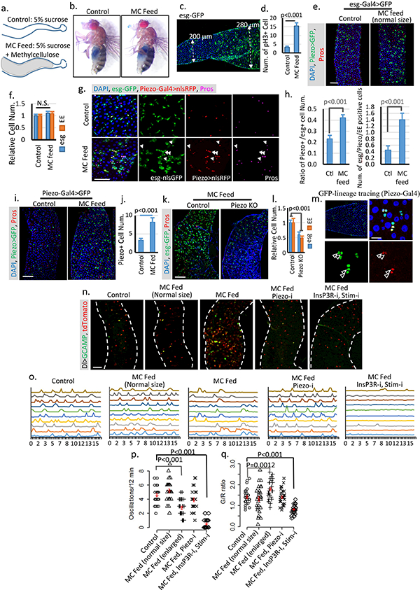
Extended Data Figure 8. Over-feeding triggers stem cell proliferation and EE increase.

a. Schematic illustration of fly midguts from control (5% sucrose) or MC (5% sucrose + 10% Methylcellulose) fed flies. b. “Smurf” assay of flies fed on both control and MC food shows no damage of gut integrity. Two independent replicates showed similar results. c,d. Image of a midgut feed on MC food. The cell proliferation phenotype is associated with midgut diameter increase but not food content. Data are collected from 23 midgut areas from two independent experiments for each condition. e,f. Midguts from flies fed on MC food with no increase of gut diameter shows no phenotype compared with control. Data are collected from 31 regions (control) and 28 regions (MC feed) from three independent experiments. g,h. Feeding-induced cell proliferation produces more Piezo+ cells, which differentiate into EEs. All newborn EEs are indicated by white arrowheads. Data are collected from 27 areas from 2 independent experiments for each condition. i,j, Feeding-induced midgut enlargement triggers a significant increase in EP/Piezo+ cell number. Data are collected from n=30 (control) and n=32 (MC feed) midgut areas from two independent replicates. k,l. Feeding-trigged stem cell proliferation and EE increase are blocked in Piezo null mutant. Data are collected from n=27 (control) and n=32 (PiezoKO) midgut areas from two independent replicates. P-values for both esg and EE are smaller than 0.001. m. Linage tracing experiment (using Piezo-Gal4) under overfed condition shows a significant increase in cell number (2–3) in the same cluster compared to tracing result under control condition, suggesting that either more Piezo cells were created from ISCs or more Piezo+ cells divide to create more progeny. Cells positive for both GFP and Pros are indicated by arrowheads. n, Images of live midguts from the following conditions/genotypes: control, MC fed without midgut diameter increase (normal size), MC fed with enlarged midgut diameter, MC fed with PiezoRNAi and enlarged midgut diameter, and MC fed with InsP3RRNAi + StimRNAi and enlarged midgut diameter. o. Representative traces of Ca2+ oscillations in Dl+ stem cells of flies from indicated treatment/genotypes. Data are collected from 3 independent experiments for each genotype/condition. p,q. Ca2+ oscillation frequency and GCaMP/RFP intensity ratio of 30 cells from 3 individual guts for each genotype are plotted. Mean ± s.e.m. is displayed in red. Enlarged midgut fed on MC food shows reduced Ca2+ oscillation frequency but increased average cytosolic Ca2+ level. MC food alone does not trigger any significant change of Ca2+ activity. Knocking-down either Piezo or both Stim and InsP3R blocks this feeding-induced increase of cytosolic Ca2+. Knocking-down InsP3R or Stim alone has no significant effect on cytosolic Ca2+ (Data not shown), which is probably due to the reduced expression level of Dl-Gal4 compared with esg-Gal4. The change of Ca2+ activity in MC-fed enlarged midguts is similar to some cells in the Bleomycin damaged midguts (Extended Data Fig. 6 f,g). However, the majority of cells from MC-fed enlarged midguts still oscillate, which is different from stem cells in Bleomycin-treated midguts in which a large portion of cells maintain a constant high level of Ca2+ (Extended Data Fig. 6 f,g). Data are indicated as mean + s.e.m. values. P-values are calculated from two-tailed Student t-test with unequal variance. Scale bar: e,i,k,n, 50 μm; g, 25 μm; m, 10 μm.
