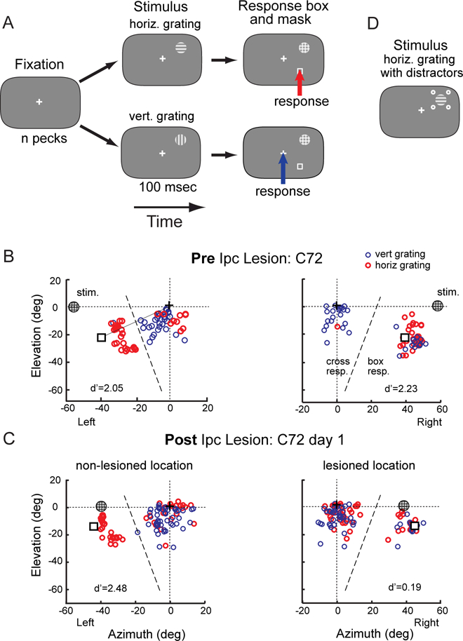
Figure 1. Orientation discrimination task and spatial patterns of peck responses.

(A) The sequence of stimuli for the single-location and mirror-locations protocols are shown as a time series from left to right. The chicken was rewarded with brief access to food for pecking within 15º of the box following the horizontal grid (red arrow) or of the cross following the vertical grating (blue arrow). The visual stimuli and locations are not drawn to scale. (B) The spatial pattern of peck responses on the touch-sensitive computer screen for a single test session from C72 before an Ipc lesion (baseline). Responses to the horizontal grating are shown in red, and responses to the vertical grating in blue. The locations of the grating, response box and cross on the screen are indicated. This bird was tested with the mirror-locations protocol (Methods), in which two grating locations at mirror symmetrical positions (circled plaids at left and right 58º, 0º elevation) were randomly interleaved. The dashed line indicating the boundary used to define box responses and cross responses was drawn perpendicular to the line connecting the cross to the box (solid line). The d’ values for this particular test session are indicated. (C) Peck responses recorded on day 1 after the Ipc lesion. The lesioned (right) and non-lesioned (left) locations (at left and right 39º, 0º elevation) were tested on randomly interleaved trials. (D) Grating surrounded by distractors, used to test birds in group 2. The contrast of the four distractor dots was varied randomly across trials. See also Figure S1.
