
Figure 4. Histological reconstructions of OT lesion sites.
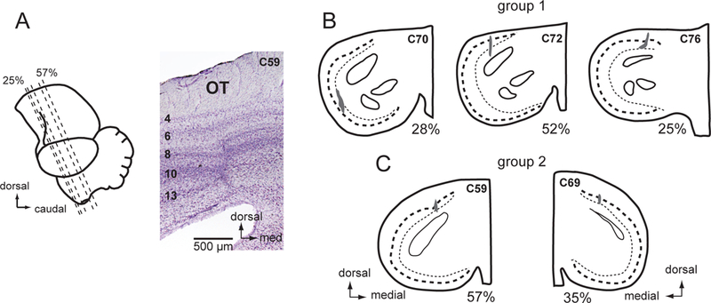
(A) Lateral view of the chicken brain showing the plane of section and a cresyl violet stained transverse section through the center of the OT lesion in C59. Numbers on the left identify the OT layers. (B) OT lesions in the group 1 birds, C70, C72, and C76. Camera lucida drawings of cresyl violet stained transverse sections. The dark shading indicates the extent of overt scaring. Dashed line: layer 10; dotted line: layer 13. The % rostrocaudal location of each section is defined in (A).(C) OT lesions in the group 2 birds, C59 and C69.