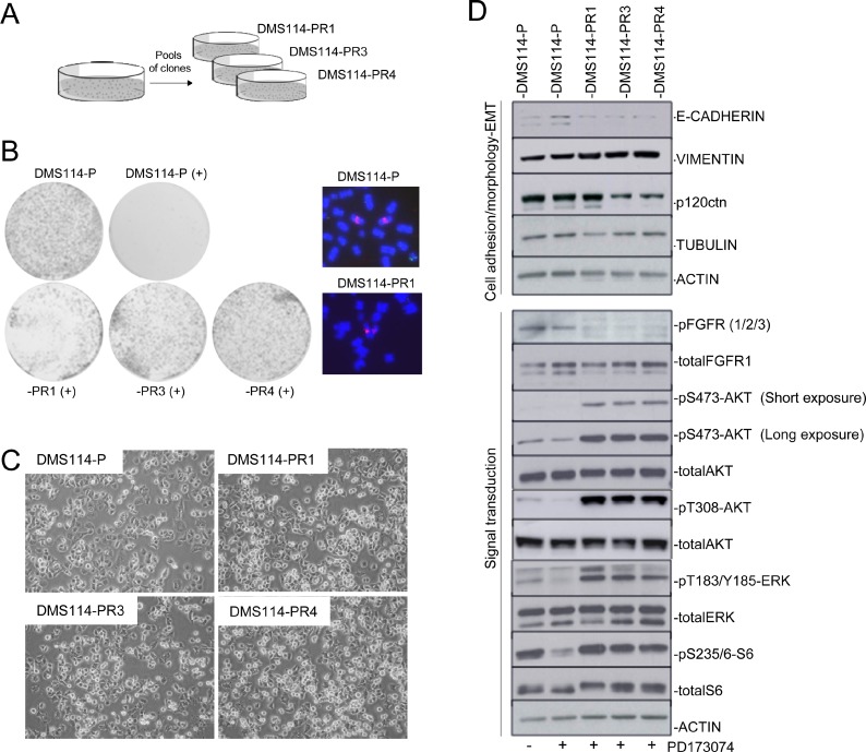
Figure 1. Generation of AR to FGFR1 inhibitors involves de novo activation of AKT and ERK.
(A) Description of the DMS114-R cells generated. (B) Left panel: Colony formation assay for cell-growth inhibition upon administering PD173074 treatment to the DMS114 parental (DMS114-P) and to the different DMS114-R cells. Right panel: Examples of metaphase nuclei from DMS114-P cells and the indicated resistant cells at the FGFR1 gene (probes in red). Control probe in green. (C) Phase contrast images showing the cell morphology of the indicated DMS114 cells. (D) Western blot of the indicated proteins in DMS114-P and DMS114-R cells. In the case of DMS114-P, extracts with (+) and without (−) treatment with the PD173074 inhibitor (1 µM) are shown. The upper panels indicate the levels of different proteins related to cell adhesion and morphology. The lower panels show the phosphorylation levels of proteins involved in signal transduction pathways. ACTIN, total protein loading controls.

