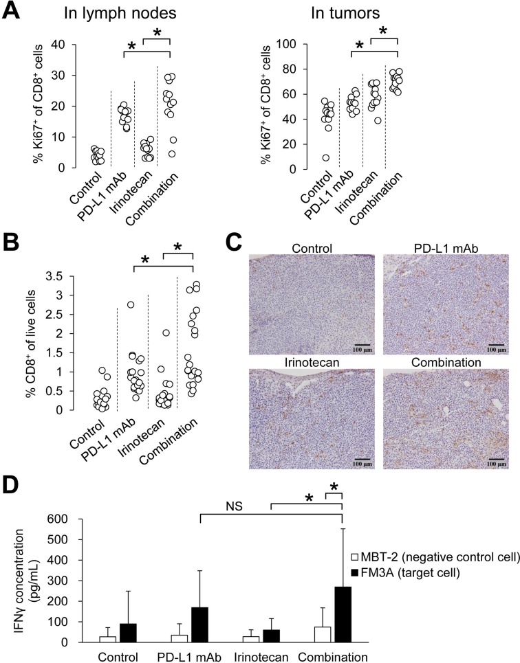
Figure 3. Combination of irinotecan plus PD-L1 mAb enhanced proliferation of CD8+ T cells and increased number of tumor-infiltrating CD8+ T cells without loss of PD-L1 blockade-induced tumor-specific lymphocyte response.
(A) Proliferation of CD8+ T cells in lymph nodes and tumors on Day 8 (n = 12/group). (B) Percentage of CD8+ T cells in tumor at the end point of the study (Day 19) (n = 19–21/group). CD8+ T cells were determined by flow cytometric analysis. (C) Infiltration of CD8+ T cells in tumors was determined by CD8α immunostaining in tumor tissue at the end point of the study (Day 19). (D) Secretion of IFNγ after specific stimulation of lymphocytes by co-culturing with tumor cells (n = 21/group). IFNγ was quantified by ELISA. Data are shown as the mean + SD. Statistical analysis used Wilcoxon rank sum test and the method of Holm.

