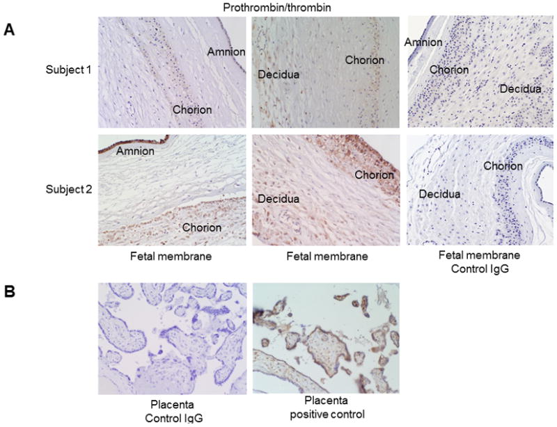
Figure 1. Prothrombin/thrombin staining in fetal membranes.

(A) Prothrombin/thrombin protein was present in the amnion, chorion, and decidua layers of the fetal membranes. Prothrombin/thrombin immunostaining intensity in Subject 2 was much higher than in Subject 1, which represented the variable expression of this protein among term fetal membranes. Negative control fetal membranes were stained negative. (B) Negative control and positive control of prothrombin/thrombin immunostaining in placenta tissues. Thrombin/prothrombin staining in placentas was in syncytiotrophoblasts and fetal vessels.
