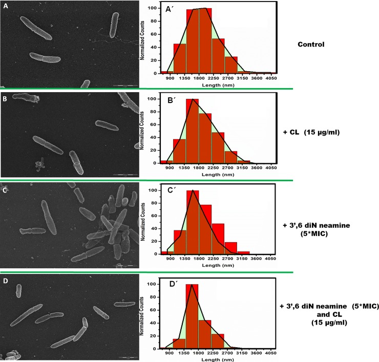
Fig 7.
Scanning electron microscopy images and length distribution of P. aeruginosa incubated without (A) or in presence of cardiolipin at 15 μg/ml in the medium before sample preparation (B), 5 *MIC 3',6-dinonylneamine (C) and with both cardiolipin at 15 μg/ml and 5 *MIC 3',6-dinonylneamine (D) together before sample preparation. Mean bacterial length was 1915 nm, 1877 nm, 1649 nm and 1767 nm, respectively with SD = 35 nm. Distribution are shown, overlaid onto the distribution profile (in red). Time of incubation was 1 hour and at least 200 bacteria were monitored. Scale bars correspond to 2 μm.

