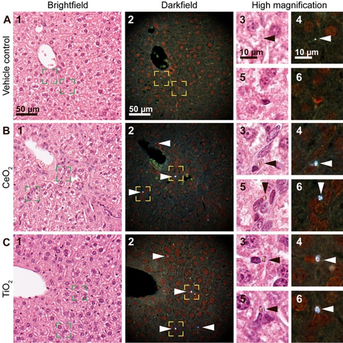
Fig 4. Brightfield (1) and enhanced darkfield (2) microscopy images of H&E stained liver tissue.
(A) From intratracheally instilled mice that received a control vehicle, (B) 162 μg/animal of CeO2 or (C) or TiO2 NPs at 180 days post-exposure. In the liver sections from mice exposed to CeO2 and TiO2 foreign material aggregates were observed using enhanced darkfield microscopy, mainly in sinusoids and often close to small nuclei (B and C, respectively, 2, 4 and 6, white arrowheads). The aggregates were not detectable in brightfield at 40x magnification (B1, C1), but exhibited a brownish appearance at 100x magnification (B and C, 3 and 5, black arrowheads). Appearance of a typical artefact is shown (A, 3–4, arrowheads). Panels 3–6 correspond to the rectangular zones in panel 1–2 captured at higher magnification.

