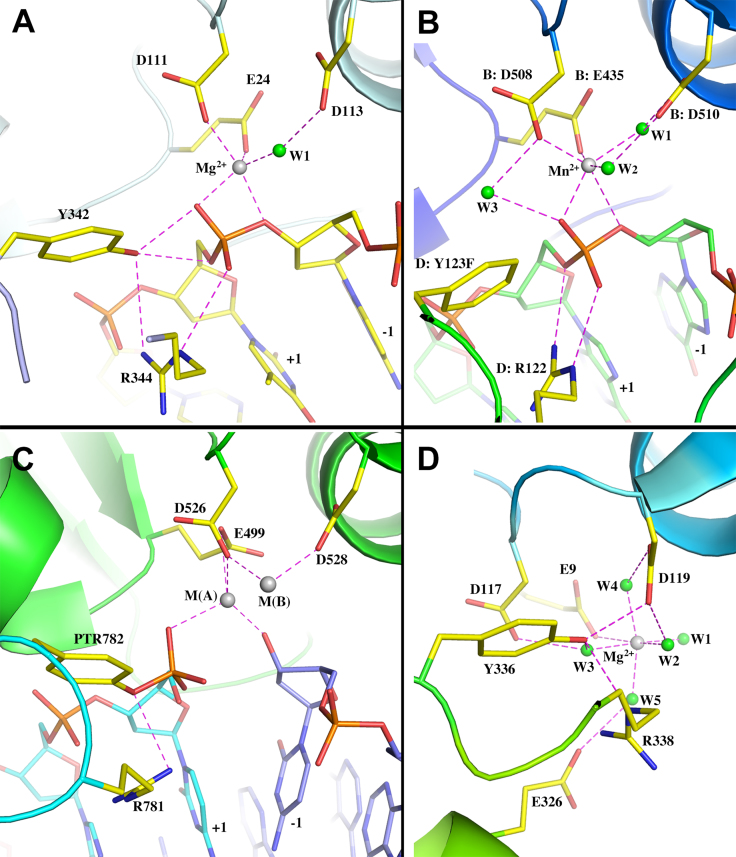
Figure 4.
Comparison of metal-binding sites of type IA and IIA topoisomerases. (A) Ribbon diagram of Mg2+ binding site of MtTOP1-704t/MTS2-13/Mg (PDB code: 6CQ2). (B) Ribbon diagram of the Mn2+ binding site of S. aureus type IIA topoisomerase complex with inhibitor and DNA (PDB code: 2XCS). Residues are labeled based on the numberings in PDB file. Waters are renumbered for convenience. (C) Ribbon diagram of the S. cerevisiae type IIA topoisomerase covalent complex with two Zn2+ binding sites (PDB code: 3L4K). Residues are labeled based on the numberings in PDB file. (D) Ribbon diagram of the active site of human topoisomerase IIIα (PDB code: 5GVC). Residues are labeled based on the numberings in PDB file. Waters are renumbered for convenience.

