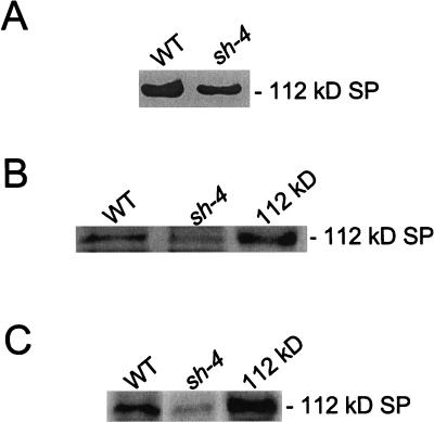
Figure 5.
Levels of the 112-kD stromal SP in the shrunken-4 mutant. A, Samples (30 μg) of the endosperm fraction from wild-type (WT) and the shrunken-4 (sh-4) mutant were subjected to native PAGE in the presence of 24 μm glycogen. Following electrophoresis, SP activity was measured by iodine staining. B, The isolated 112-kD protein (0.1 μg) and samples (60 μg) of the endosperm fraction from wild-type (WT) and the shrunken-4 (sh-4) mutant were subjected to SDS-PAGE followed by Coomassie Blue staining. C, The isolated 112-kD protein (0.1 μg) and samples (30 μg) of the endosperm fraction from wild-type and the shrunken-4 mutant were subjected to immunoblot analysis using anti-SP antibodies. A portion of the polyacrylamide gels (A and B) and the immunoblot (C) is shown, and the position of the 112-kD stromal SP is indicated in the figure. The data shown in A through C is representative of two independent experiments.

