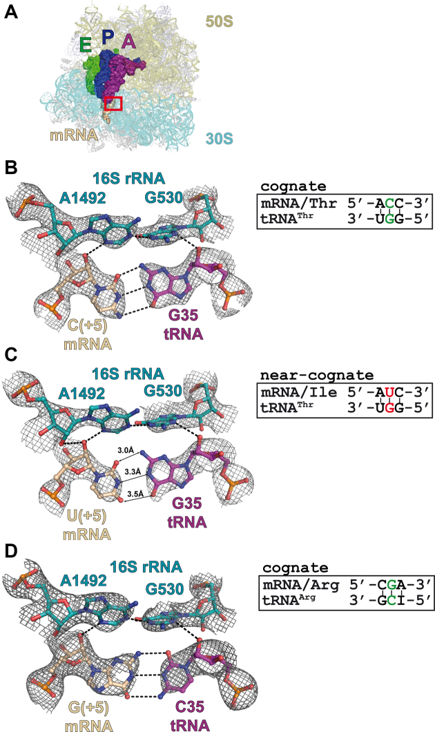
Figure 1.
Complexes of the Thermus thermophilus 70S ribosome with mRNA and tRNA in the A-site. (A) Overview of the complex structure. The red frame marks the decoding center. 16S rRNA is shown in teal, 23S rRNA—in olive, A-site tRNA—in purple, P-site tRNA—in blue, E-site tRNA—in green and mRNA—in wheat. Views of the second codon–anticodon base pair in the cognate (B) and near-cognate (C) tRNAThrGGU complexes. (D) Second codon–anticodon base pair in the cognate tRNAArgICG complex. Putative hydrogen bonds (distances ≤3.3 Å) are shown as dashed lines and interatomic distances for the mismatched pair are indicated. 2Fo–Fc electron density maps are contoured at 1.2 σ.

