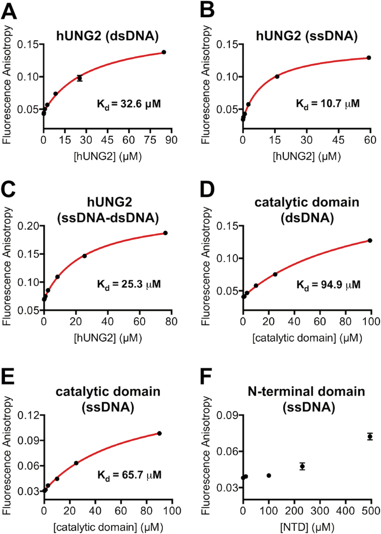
Figure 4.
Fluorescence anisotropy binding assays. Except for panel F, the buffer consisted of 25 mM HEPES–NaOH (pH 7.4), 10% glycerol, 100 mM NaCl, 1 mM MgCl2, 1 mM DTT, and 0.01% Triton X-100. In all assays, the fluorescein-labeled DNA was 50 nM. (A) Binding of hUNG2 to a 29 bp duplex. (B) Binding of hUNG2 to a 29 nt ssDNA. (C) Binding of hUNG2 to a hybrid ssDNA–dsDNA duplex containing a 29 nt 5′ ssDNA overhang and a 29 bp duplex region. (D) Binding of the catalytic domain to a 29 bp duplex. (E) Binding of the catalytic domain to a 29 nt ssDNA. (F) Binding of the isolated N-terminal domain (residues 1–91) to a 29 nt ssDNA in a buffer with 20 mM NaCl.

